
Incubator Accessories

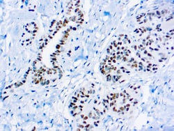
Diagnostic Biosystems WILMS TUMOR 1 PROTEIN WT1 Wilm’s Tumor 1, WT1
Wilm’s Tumor (WT1), is a gene involved in the induction of Wilm’s Tumor, a sporadic and familial pediatric kidney tumor. Wilm’stumor is associated with mutations of WT1, a zinc-finger transcription factor that is essential for the development of the metanephric kidney and the urogenitalsystem. The WT1 gene is normally expressed in fetal kidney and mesothelium, and its expression has been suggested as a marker for Wilm’s tumor and mesothelioma. WT1 has been identified in proliferative mesothelial cells, malignant mesothelioma, ovarian carcinoma, gonadoblastoma, nephroblastoma, and desmoplastic small round cell tumor.

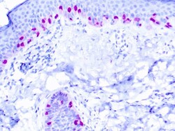
Diagnostic Biosystems MICROPHTHALMIA Microphthalmia Transcription Factor, MiTF
This antibody reacts with a 52-56 kDa protein known as microphthalmia (Mi). This antibody reacts with both melanocytic and non-melanocytic isoforms of Mi. Mi is a basic helix-loop-helix-leucine zipper (b-HLH-ZIP) transcription factor implicated in pigmentation, mast cells and bone development. There are two known isoforms of Mi differing by 66 amino acids at the amino terminus.
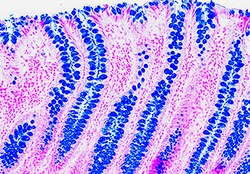
Diagnostic Biosystems ALCIAN BLUE PH 25 Alcian Blue, pH 2.5
The Alcian Blue (pH 2.5) Stain Kit is intended for use in the histological visualization of sulfated and carboxylated acid mucopolysaccharides and sulfated and carboxylated sialomucins (glycoproteins).
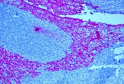
Diagnostic Biosystems UnoVue™ Mouse AP Detection Reagent
UnoVue Mouse AP Detection Reagent is a non-biotin one-step detection reagent suitable for demonstrating antigens in formalin-fixed paraffin-embedded tissues and frozen sections. The UnoVue detection reagents may also be used with blood smears, cytosmears, and cell preparations. UnoVue detection reagents have been developed by directly labeling anti-mouse immunoglobulins with enzymes using a proprietary tandem hyperlabelling technology. This ensures consistent and reproducible immunodetection of mouse antibodies against nuclear, cytoplasmic and membrane antigens in different types of tissues. The single step UnoVue Detection Reagent enables faster staining procedures than traditional two-step methods using biotin and avidin/streptavidin conjugates, with significantly lower background. UnoVue Mouse AP Detection Reagent is suitable for use with mouse all mouse antibodies, both monoclonal and polyclonal.
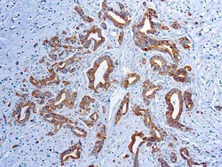
Diagnostic Biosystems VASCULAR ENDOTHELIAL GROWTH FA Vascular Endothelial Growth Factor (VEGF) (VG1)
This antibody is specific to a cluster of proteins between 19-22 kDa (reduced). VEGF is a homodimeric disulfide-linked glycoprotein involved in angiogenesis, which promotes tumor progression and metastasis. It exhibits potent mitogenic and permeability-inducing properties specific for the vascular endothelium.
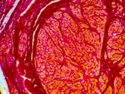
Diagnostic Biosystems PICRO SIRIUS RED STAIN KIT Picro Sirius Red Stain Kit
The Picro-Sirius Red Stain Kit (For Collagen) is intended for use in the histological visualization of collagen I and III fibers in addition to muscle in tissue sections.
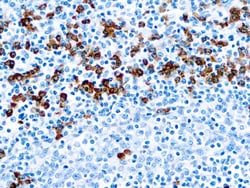
Diagnostic Biosystems IGG IgG
This antibody reacts with γ-chains of human IgG. Nonspecific antibodies have been removed by solid-phase adsorption.

Diagnostic Biosystems ALK PHOS ENHANCER Alkaline Phosphatase Enhancer
This product is used as a signal enhancer in conjunction with alkaline phosphatase based immunostaining systems.\n\nAlkaline Phosphatase, an enzyme derived from bovine intestinal mucosa, is often used as a label for in situ hybridization, immunohistochemistry, Southern and Northern hybridization and DNA sequencing. Detection of this enzyme requires the generation of an insoluble colored reaction end product. Stronger the signal, better the staining. DBS introduces a new stable solution to increase the signal generated by alkaline phosphatase several fold. A single treatment of tissue sections with enhancer before the treatment with substrate/chromogen enables the visualization of difficult to localize antigens. This enhancer works well with Fast Red but can be used with BCIP/NBT and BCIP/INT also. Specimens stained with the above substrates/chromogens can not be dehydrated in ethanol and hence should be mounted in aqueous based mounting medium like C/C Mount (DBS catalog # K002).
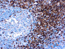
Diagnostic Biosystems CD4 CD4 (EP204)
CD4 is glycoprotein found on the surface of immune cells such as T helper cells, monocytes, macrophages, and dendritic cells. It is co-receptor that assists the T-cell receptor (TCR) with antigen-presenting cell and also interacts directly with MHC class II molecules of the surface of antigenpresenting cells. Most mature T-cell lymphomas are CD4 positive with the exception of aggressive NK-cell leukemia and extranodal NK/T-cell lymphoma. CD4 plays important role in the classification of lymphocytes in inflammatory legions and malignant lymphomas.
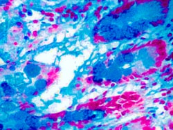
Diagnostic Biosystems ALCIAN BLUE PH 10 Alcian Blue, pH 1.0
This kit stains strongly sulfated mucosubstances. Strongly sulfated mucosub-stances stain blue in paraffin tissue sections, while the nuclei stain pink to red and the cytoplasm and background stain pale pink.
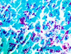
Diagnostic Biosystems AFB AFB
This kit stains acid-fast bacteria and tubercle bacilli of Mycobacterium genus in paraffin tissue sections. Tubercle bacilli and other acid-fast bacteria stain bright red and the background stains light green.
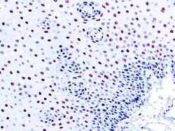
Diagnostic Biosystems ERCC1 ERCC1
This antibody reacts with a 33-36 kDa protein known as ERCC1 (excision repair cross complementing) polypeptide. ERCC1 is required for nucleotide excision repair of damaged DNA and is homologous to RAD10. In mammalian cells, XPG cleaves 3’ of the DNA lesion while ERCC1-XPF complex makes the 5’ incision.
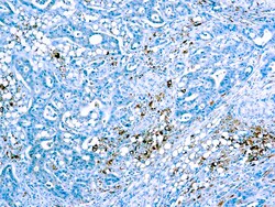
Diagnostic Biosystems CATHEPSIN D Cathepsin D
Cathepsin D is an estrogen-regulated protein associated with tissue breakdown. Levels of cathepsin D have been positively correlated with recurring breast cancers of both node-negative and node-positive types. Additionally cathepsin D has been associated with amyloid formation in Alzheimer's plaques. As cathepsin D activity is increased by cigarette smoke, the enzyme may contribute to lung tissue damage in smokers.
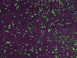
Diagnostic Biosystems LAMBDA LIGHT CHAIN-FITC Lambda Light Chain-FITC
Traces of contaminating antibodies have been removed by solid phase absorption. Only the lambda precipitation line appeared in cross immunoelectrophoresis. This antibody is well suited for tissue section staining but may also be used for other applications.
